Wildlife in Texas - Types of Texan Animals


This Lady Lizard Can Reproduce Without A Mate
This story appears in the November 2017 issue of National Geographic magazine.The Komodo dragon, the largest species of lizard now alive, can grow to 10 feet long and nearly 200 pounds. A muscular carnivore armed with sharp teeth, Varanus komodoensis dines on prey as large as deer, wild boar, and water buffalo.
As formidable as the Komodo dragon seems, it's very much at risk. On seven Indonesian islands that are the only places the species lives in the wild, humans burn its habitat to clear land and poach animals on which it preys. Even on land protected as Komodo National Park, officials reported just 3,013 Komodo dragons in 2016, down from 3,222 in 2013.
When her species needs replenishing, what's a mother dragon to do? She can reproduce the old-fashioned way, by mating with a male and laying eggs. Or she can lay eggs without having mated, through a sort of virgin birth process called parthenogenesis.
In 2006 at England's Chester Zoo, a female named Flora, who'd had no male contact, laid a clutch of viable eggs that tests showed had only her DNA. That was the first confirmation of parthenogenesis in captive Komodo dragons; scientists now believe it "happens very often," says Gerardo Garcia, Chester Zoo's curator of lower vertebrates and invertebrates.
How does it work? In humans, males have both male and female sex chromosomes. In Komodo dragons, females do—so Flora had within her the genetic materials needed for embryos to develop. This self-fertilization yields offspring that are "absolutely healthy," Garcia says—but every one is male.
Being able to reproduce both sexually and asexually gives the dragons an evolutionary edge, Garcia says. If no mate is handy, a female can bear sons parthenogenetically—and when they're older, they can be her mates. "It's not ideal" for keeping the gene pool diverse, he says. But it's a way for the species to continue.
Habitat/range: Tropical-forest edge and grasslands of seven Indonesian islands
Conservation status: IUCN assesses it as vulnerable.
Other facts: One of about 80 species of monitor lizards, Komodo dragons have existed for perhaps 3.8 million years, fossils suggest.
Australian Lizards Have Evolved Resistance To Deadly Venom, But Some Snakes 'fire Back'
Bryan Fry recalls watching a battle unfold in the Australian outback between two arch-enemies: the sand goanna and the venomous western brown snake.
"The brown snake was biting it and really drawing blood, but the sand goanna was totally unaffected and ended up slurping down the brown snake," he said.
The two reptiles were entangled in an evolutionary arms race.
"We know that the big species of goanna feed a lot on venomous snakes, while the smaller species are obviously small enough to be vulnerable to a venomous snake as far as being potential prey," Professor Fry, a toxicologist at the University of Queensland, said.
In this case, the brown snake drew first blood, but evolution equipped the sand goanna with the ability to combat the snake's highly toxic venom.
Professor Fry with a desert spotted monitor (Varanus panoptes rubidus), commonly known as a sand goanna, in Western Australia. (Supplied: Bryan Fry)
Professor Fry is part of a team of researchers that has been investigating how some species of lizards have evolved to survive being bitten by some of the world's deadliest snakes, and why others become a tasty meal.
Their research, which was published in the International journal of Molecular Sciences, focused on the interactions between elapids, a family of highly venomous snakes, and varanids, a family of lizards.
Elapids include death adders and cobras, while goannas and Komodo dragons number among the varanids.
The researchers wanted to compare how the reptiles evolved in response to being both predators and prey, Professor Fry said.
"It was really interesting to see how it all shook up, but we never expected it to be so incredibly complex."
Evolutionary arms raceElapid snake venom contains neurotoxins which interfere with the transmission of nerve impulses by blocking chemical receptors on muscle fibres so they can't contract, leading to paralysis and death.
To understand how different species of lizards responded to snake bites, researchers applied venom to tiny patches of tissue, sourced from dead lizards in museum collections across Australia.
The study found that most large varanid lizards, such as goannas, have inherited neurotoxin resistance to the venom – a trait Professor Fry said was possibly connected to their predatory lifestyle.
"The key is that the lizards are evolving resistance to alpha-neurotoxins, which is a toxin type that is shared across Australian elapid snakes," he said.
Professor Fry said they discovered multiple mutations in the receptors that were responsible for the lizards' resistance, which were passed down to their offspring and future generations.
But not all lizards "shield themselves in the same way", he said.
The study found that two lineages of giant varanids – the Komodo dragon and the perentie – had reduced chemical resistance to the venom, but would use their physical build to armour themselves against the snakes.
Komodo dragons (Varanus komodoensis) are the largest living lizard species in the world. Full-grown adults can reach up to 3m in length and weigh more than 135kg. (Supplied: Adelaide Zoo)
"Their thick, bone-filled scales are enough to protect them against snakebite, while their large teeth are used to quickly dismember the fettuccine-like snakes," Professor Fry said.
"Obviously, that's not going to be enough to protect them against a bite, but as they become bigger and bigger, it becomes like chain mail. Eventually, it's going to be to the point where the snakes aren't even going to be able to penetrate [their skin]."
This indicated that size and mechanical defences may reduce the need for chemical resistance – "in essence, evolution has favoured brawn over biochemistry," he said.
It's not just size that countsResearchers also found dwarf varanids such as tree monitor lizards lost the resistance as they evolved to live above the ground and away from predators.
Other dwarf lineages such as the rusty desert monitor and the short-tailed pygmy monitor re-evolved the resistance when they came down from the trees and evolved to become burrowers.
Two dwarf goannas, a rusty desert monitor (Varanus eremius) on the left and short-tailed pygmy goanna (Varanus brevicauda) on the right. They typically spent most of their time hiding under rocks or within burrows.(Picture: Bryan Fry)
Uthpala Chandrasekara, a PhD candidate from the University of Queensland who completed the research work as part of her thesis, said the results illustrated the dynamic nature of the evolutionary process – and why researchers framed their observations as "a Russian doll of resistance."
"What this describes is how lizards have evolved their immunity to venom in a complex and layered manner, similar to how these Russian dolls fit one inside another," she said.
"Specific traits can emerge, be lost and re-emerge across different varanid species in response to the changing ecological and biological processes."
The results also suggested the evolutionary battle doesn't always head in one direction.
"Once the lizards evolve, the snakes fire back with even more potent toxins," Ms Chandrasekara said.
"It's a biological arms race where the only constant is change."
The study discovered that some Australian elapid snakes, such as the death adder, have developed highly potent venoms, which are more effective at counteracting the lizards' defences.
Some death adders (Acanthophis) have developed more potent toxins specifically targeting resistant varanid lizards.(Supplied: Luke Allen)
When the lizards develop resistance, this then puts a reciprocal pressure on the snakes, "for their venom to become more potent, or more selective, or more efficient because otherwise they're not going to feed," Professor Fry explained.
"It's a classic back and forth, where a change in the venom puts a pressure on the prey, but a change in the prey susceptibility puts pressure back on the venom."
The only way to win, he said, was to not play the game.
"The little lizards that are up in the trees are the true winners because they're completely decoupled from the entire arms race now."
'Deeply fascinating'Timothy Jackson, co-head of the Australian Venom Research Unit at the University of Melbourne, said the study was "deeply fascinating" and increased our knowledge of how toxin resistance in varanid lizards worked at the molecular level.
ABC Science on YouTube Read more"It is also a strong contribution to the literature on the potential for evolution of venom resistance," Dr Jackson, an evolutionary toxicologist with a focus on reptile venom, said.
But he said there were still questions to answer about how ecological factors such as feeding and defence contributed to evolution.
"As is typically the case in scientific investigations, interesting patterns are observed and there is a reasonable desire to conjecture an explanation for those patterns.
"The specific explanations conjectured in terms of feeding and defensive ecology require further investigation."
He clarified that it was important that "evolutionary biologists should make such conjectures".
"Imagining the encounter between predator and prey and the attendant selection pressures is part of what fuels our desire to investigate these fascinating systems."
Get all the latest science stories from across the ABC.
The Impact Of Apicobasal Ridges On Dental Load-bearing Capacity In Aquatic-feeding Predatory Amniotes
IntroductionGeological time has borne witness to many reinvasions of the aquatic environment by lineages of terrestrial tetrapods (Fish Reference Fish2016). These reinvasions were dependent upon a variety of evolutionary shifts in morphological and behavioral traits (Fish Reference Fish2016; Houssaye and Fish Reference Houssaye and Fish2016). The return to aquatic environments placed immense selective pressure on the terrestrial Bauplan, resulting in widespread convergence of morphological traits between secondarily aquatic tetrapods (Howell Reference Howell1930; Braun and Reif Reference Braun and Reif1985; Thewissen and Nummela Reference Thewissen, Nummela, Thewissen and Nummela2008; Houssaye and Fish Reference Houssaye and Fish2016). Although recent research into aquatic-feeding predators has focused on the dietary significance of tooth shape (Lukeneder and Zverkov Reference Lukeneder and Zverkov2020; Sulcova et al. Reference Sulcova, Zahradnicek, Dumkova, Dosedelova, Krivanek, Hampl and Kavkova2020; Fischer et al. Reference Fischer, Bennion, Foffa, MacLaren, McCurry, Melstrom and Bardet2022), little is known about the function of crown surface morphology. This gap is particularly evident in the case of apicobasal ridges, longitudinal enamel ridges present in several clades of secondarily aquatic amniotes (McCurry et al. Reference McCurry, Evans, Fitzgerald, McHenry, Bevitt and Pyenson2019) (Fig. 1). The structural makeup of these ridges varies between taxa; cross-sectional analysis by McCurry et al. (Reference McCurry, Evans, Fitzgerald, McHenry, Bevitt and Pyenson2019) demonstrates that in plesiosaurs, the ridges do not extend into the dentine, resulting in thicker enamel at their peaks, which may be due to differential growth rates between enamel types (Sander Reference Sander1999, Reference Sander, Teaford, Smith and Ferguson2000). In other clades, including spinosaurids, odontocetes, and crocodilians, the intersection between dentine and enamel extends parallel to the outer surface of the ridges (McCurry et al. Reference McCurry, Evans, Fitzgerald, McHenry, Bevitt and Pyenson2019). In ichthyosaurs, the ridges mirror an internal dentine folding known as plicidentine, which begins within the crown and extends toward the root (Maxwell et al. Reference Maxwell, Caldwell and Lamoureux2012; McCurry et al. Reference McCurry, Evans, Fitzgerald, McHenry, Bevitt and Pyenson2019). These disparate structural morphotypes support a convergent result, implying distinct amelogenetic pathways to an analogous trait (Sander Reference Sander1999; McCurry et al. Reference McCurry, Evans, Fitzgerald, McHenry, Bevitt and Pyenson2019).
Figure 1. Bauplan silhouettes and tooth faces bearing apicobasal ridges from members of six major aquatic-feeding amniote clades. A, Pliosaurus brachydeirus (Plesiosauria) (ROM 5596). B, Pervushovisaurus campylodon (Ichthyosauria) (ROM 00334 A). C, Deinosuchus rugosus (Crocodylia) (USNM 5351). D, Tylosaurus proriger (Mosasauridae) (USNM 3885). E, Spinosaurus sp. (Spinosauridae) (ROM 64659). F, Aetiocetus cotylalveus (Odontoceti) (USNM 25210 Tooth 1). Arrows indicate apicobasal ridges.
Apicobasal ridge morphologies vary in shape, size, and arrangement. The abundance of morphological variation, even within some lower-ranked taxa, suggests that—like crown shape (Massare Reference Massare1987)—ridge morphologies are related to diet rather than clade, although species specificity does occur (Benson et al. Reference Benson, Fitzgerald, Rich and Vickers-Rich2013; Zverkov et al. Reference Zverkov, Fischer, Madzia and Benson2018). One example of this species specificity is the common characterization of plesiosaur teeth by widely spaced, distinct ridges on a smooth surface, with an often trihedral tooth shape (Benson et al. Reference Benson, Fitzgerald, Rich and Vickers-Rich2013; Zverkov et al. Reference Zverkov, Fischer, Madzia and Benson2018).
Some specimens with ridges around the entire crown circumference, such as some Late Cretaceous material previously attributed to Polyptychodon, display density that differs between tooth faces, with lingual ridges clustering more closely than buccal ridges (Madzia Reference Madzia2016). In taxa with trihedral or subtrihedral crown cross sections, ridges may be present on only one or two of the faces, typically excluding the buccal face (Fischer et al. Reference Fischer, Arkhangelsky, Stenshin, Uspensky, Zverkov and Benson2015). These taxa also often trend toward an increased clustering of ridges on the lingual surface (Taylor Reference Taylor1992; Buffetaut Reference Buffetaut2013; Richter et al. Reference Richter, Mudroch and Buckley2013).
The ridges themselves can be high or low relief, varying within individual teeth as well as between species, but remain visually distinct from smooth enamel (Buffetaut Reference Buffetaut2013). Coverage of the apicobasal length is also variable; across taxa, ridges taper off or anastomose anywhere from a few millimeters above the base to directly below the apex (Massare Reference Massare1987; Young et al. Reference Young, Steel, Brusatte, Foffa and Lepage2014; Madzia Reference Madzia2016).
The strength hypothesis is among the most frequently cited hypotheses aiming to explain the function of apicobasal ridges in aquatic feeding strategies. This hypothesis focuses upon the potential for the ridges to strengthen the tooth crown (Preuschoft et al. Reference Preuschoft, Reif and Müller1974; Vaeth et al. Reference Vaeth, Rossman and Shoop1985; Young and Kardong Reference Young and Kardong1996; Schulp Reference Schulp2005; Young et al. Reference Young, Steel, Brusatte, Foffa and Lepage2014; Zverkov et al. Reference Zverkov, Fischer, Madzia and Benson2018), possibly by channeling pressure toward its base and the "valleys" between ridges (Preuschoft et al. Reference Preuschoft, Reif and Müller1974; Rieppel and Labhardt Reference Rieppel and Labhardt1979; Sander Reference Sander1999) or by increasing the second moment of area (I), an engineering principle that quantifies the rigidity of a shape and would enable the ridges to act comparably to corrugations in a metal sheet by increasing the load-bearing surface area (Khalid et al., Reference Khalid, Chan, Sahari and Hamouda2004). It should be noted, however, that the latter comparison is somewhat confounded by the addition of dentine below the enamel ridges, increasing I, while metal sheets generally bear no additional material below their corrugations. As the strength hypothesis is only one of several plausible hypotheses, the remainder of which include improving grip on slippery prey and enhancing puncture efficiency (McCurry et al. Reference McCurry, Evans, Fitzgerald, McHenry, Bevitt and Pyenson2019; Crofts et al. Reference Crofts, Smith and Anderson2020), testing is required to better understand the physical capabilities of the ridges and any strength advantages they may have conferred.
Here we aim to assess the impact of apicobasal ridges on the ability of the tooth crown to resist various loading conditions, through the application of finite element analysis (FEA). We compare von Mises stress responses across a range of typical apicobasal ridge morphologies, analyzing the influence of ridge shape, size, and arrangement on crown strength. Furthermore, we use comparative morphometrics to evaluate morphological variation in apicobasal ridges and determine whether morphologies are clustered by phylogeny. Although ridge morphology is relatively well documented in plesiosaurs (Benson et al. Reference Benson, Fitzgerald, Rich and Vickers-Rich2013; Fischer et al. Reference Fischer, Arkhangelsky, Stenshin, Uspensky, Zverkov and Benson2015; Madzia Reference Madzia2016; Zverkov et al. Reference Zverkov, Fischer, Madzia and Benson2018), our knowledge of trends in other taxa, such as odontocetes, ichthyosaurs, and mosasaurs, is currently limited and may be expanded through this study.
Materials and Methods Specimen Selection and ImagingForty-eight individual tooth specimens were selected to represent six major clades of aquatic-feeding amniotes (Plesiosauria, Ichthyosauria, Crocodylia, Mosasauridae, Spinosauridae, and Odontoceti) (Appendix 1). Specimen selection was informed primarily by preservation, excluding worn and damaged specimens for which a cross section could not be measured. As only one set of specimens (ROM 12809 Tooth A–F) was originally found in partial articulation, all specimens in the dataset have been treated as isolated teeth. We were unable to assign positions within the jaw for each tooth due to the prevalence of homodont dentition in marine reptiles (Ciampaglio et al. Reference Ciampaglio, Wray and Corliss2005). While position along the toothrow affects the amount of force experienced by a crown (Bourke et al. Reference Bourke, Wroe, Moreno, McHenry and Clausen2008; Cohen et al. Reference Cohen, Weller and Summers2020), it is less likely to affect potential directions of force (lateral and vertical). As this study compares responses to directional force, jaw position is unlikely to greatly affect the results. Specimens were instead selected to portray a variety of ridge and crown morphologies.
The specimens were drawn from the collections of four museums. High-resolution microcomputed tomography and neutron imaging were utilized by previous researchers to digitize the specimens (McCurry et al. Reference McCurry, Evans, Fitzgerald, McHenry, Bevitt and Pyenson2019). This process was completed using either Nikon Metrology's combined 225/450 kV microfocus X-ray and computed tomography (CT) walk-in vault system at Chesapeake Testing in Belcamp, Md., U.S.A., or the DINGO neutron beamline at Australia's Nuclear Science and Technology Organisation (ANSTO) in Lucas Heights, NSW, Australia (McCurry et al. Reference McCurry, Evans, Fitzgerald, McHenry, Bevitt and Pyenson2019). The teeth were then digitally isolated from the CT scan and exported as three-dimensional surface models in STL (standard triangular language) format by previous researchers (McCurry et al. Reference McCurry, Evans, Fitzgerald, McHenry, Bevitt and Pyenson2019).
Institutional Abbreviations KKF: Kronosaurus Korner, Richmond, QLD, Australia; NHM: Natural History Museum, London, U.K.; ROM: Royal Ontario Museum, Toronto, ONT, Canada; and USNM: National Museum of Natural History, Smithsonian Institution, Washington, D.C., U.S.A. Data CollectionThe three-dimensional models were orientated within a Cartesian coordinate system and measured in Rhino v. 6 (McNeel Reference McNeel2019). Crown height was recorded from the enamel dentine junction to the apex of the crown (Fig. 2, Appendix 2). Where the apex was incomplete, measurement concluded at the highest point of the crown to avoid overestimation of crown height, although we acknowledge this would also result in a slight underestimation within some specimens. Crown width was recorded at the widest point of the crown for both the labiolingual and mesiodistal orientations (Fig. 2, Appendix 2). For mold fossil specimens that lacked a three-dimensional crown, width was recorded only for the visible orientation.
Figure 2. Standard measurements recorded for fossilized tooth specimens. A, Crown height and position of lateral cross section at 50% of the height (dotted line). B, Crown width measurements (both mesiodistal and labiolingual). C, Apicobasal ridge height and apicobasal ridge width within a cross-section outline (not to scale). Demonstrated on a Bottosaurus sp. Tooth crown (USNM 508536).
Ridge height and width measurements were taken for 10 ridges per tooth across a lateral cross section of the crown at 50% of its height (Appendix 3). The apicobasal span of the ridges was recorded as a percentage of the total crown height (Appendix 2). If a cross section bore more than 10 ridges, 10 were randomly selected for data collection. If fewer than 10 ridges were identified, the specimen was excluded from the ridge measurement section of the study. The overall count of ridges around the circumference of the cross section were recorded, regardless of whether the tooth was included in the measurement section, to gain a holistic understanding of ridge count across all taxa. Carinae were not classified as apicobasal ridges for the purposes of this study, as they are commonly accepted to be separate structures differing in abundance and relief (Young et al. Reference Young, Steel, Brusatte, Foffa and Lepage2014; Hendrickx et al. Reference Hendrickx, Mateus and Araújo2015; McCurry et al. Reference McCurry, Evans, Fitzgerald, McHenry, Bevitt and Pyenson2019). All linear measurements were log transformed in R v. 4.0.4 (R Core Team 2021) to conform to a normal distribution and to visualize morphological differences between specimens that vary considerably in the analyzed traits.
Analysis of Ridge Morphology Linear Regression Models A range of morphological variables measured on the specimens were fit to linear regression models to investigate the dental diversity of aquatic-feeding amniotes and identify taxon-specific traits. The use of linear regression models allows an evaluation of variance in all relationships of potential interest and provides insight into isometry or allometry within the scaling mechanics of the ridges. The compared variables included relative apicobasal ridge height and width, crown height and width, and ridge count. Relative ridge measurements were included to ensure the exclusion of size bias and were calculated by dividing each tooth's average ridge height or width by the crown height or width respectively. The labiolingual and mesiodistal crown width measurements were averaged to provide a representative singular width for the regression models. All models utilizing count data were analyzed using a generalized linear model with a quasi-Poisson distribution to accommodate this type of data and variance dispersion. All regression models were conducted and plotted in R v. 4.0.4 (R Core Team 2021). Taxonomic Variance in Ridge MorphologyTo determine any significant differences between the average ridge data of the six aquatic-feeding amniote groups examined in this study, univariate Kruskal-Wallis tests were performed in R v. 4.0.4 (R Core Team 2021). These variance tests were run on data for relative crown height, relative crown width, and ridge count across all six groups. All R scripts are available in the Supplementary Material.
FEAFEA is a numerical technique enabling the digital simulation of directional force across mechanical components of complex structures (Clough Reference Clough1990; Dumont et al. Reference Dumont, Piccirillo and Grosse2005). In FEA software, the visualization of stress and strain is directed by the application of load cases and constraints in contextually relevant positions across a model (Dumont et al. Reference Dumont, Piccirillo and Grosse2005). Our study conducted FEA upon digital tooth crown models to determine the impact of apicobasal ridges upon overall crown strength. These tests utilized von Mises stress distributions to compare model performance under the simulation of various loading conditions. Von Mises stress is often incorporated into biological model analyses to establish potential failure points under a ductile model of fracture or a model in which deformation precedes breakage (Dumont et al. Reference Dumont, Grosse and Slater2009; Rajabizadeh et al. Reference Rajabizadeh, Van Wassenbergh, Mallet, Rücklin and Herrel2021). Although we acknowledge that enamel and dentine conform to a brittle pattern of fracture, thus making von Mises stress unsuitable to predict their failure points, it does offer a thorough visualization of stress distribution for these materials (Neves et al. Reference Neves, Coutinho, Alves and de Assis2015) and is frequently used as such in biomechanical analyses of teeth (Whitenack et al. Reference Whitenack, Simkins and Motta2011; Palci et al. Reference Palci, LeBlanc, Panagiotopoulou, Cleuren, Abraha, Hutchinson, Evans, Caldwell and Lee2021; Rajabizadeh et al. Reference Rajabizadeh, Van Wassenbergh, Mallet, Rücklin and Herrel2021; Pollock et al. Reference Pollock, Panagiotopoulou, Hocking and Evans2022). Thus, to enable a comparison between our results and similar biological studies, we have utilized von Mises stress to visualize our stress distribution. All FEA was completed using Strand7 v. 2.4.6.Footnote *
Model Creation The tooth crown and apicobasal ridge data were used as a foundation for the construction of a digital set of seven three-dimensional tooth crown models in Rhino v. 6 (Fig. 3; see Supplementary Material). To reflect the scope of tooth crown morphologies within the dataset, the models were based on two of the most widespread crown shapes in aquatic-feeding dentition (conical and recurved). Common variations in ridge size and shape were re-created on conical models, while variations in ridge arrangement were re-created on recurved models to replicate the range documented in recurved teeth (Taylor Reference Taylor1992; Buffetaut Reference Buffetaut2013; Richter et al. Reference Richter, Mudroch and Buckley2013). Figure 3. Comparison between select examples of ridged and smooth digital tooth models. A, Average Ridges model (ridge width = 0.663 mm, ridge height = 0.136 mm). B, No Ridges (Conical) model. C, Symmetrical Ridges model. D, No Ridges (Recurved) model. All models are in mesial view.The Average Ridges model represents the average ridge measurements, the height and width of which were applied to all models except the Large Ridges model, which instead bears both wider and taller ridges matching the maximum recorded dimensions. The Rounded Ridges model exemplifies ridges with a rounded cross section. The Lingual Clustering model bears ridges only on the recurved lingual face, while the Symmetrical Ridges model represents recurved teeth with ridges around the entire circumference.
Although many fossil specimens have ridges of varying lengths along the crown, all ridges in our models ended at 72% of the crown height (the proportional average) to ensure standardization. Control models without ridges were created for both tooth shapes to enable a comparison between identical teeth with and without ridges (Fig. 3). All models were designed to fit the average crown height and width to eliminate size bias. Although ridge shape can manifest as triangular or rounded in cross section, depending on taxon (McCurry et al. Reference McCurry, Evans, Fitzgerald, McHenry, Bevitt and Pyenson2019), triangular ridges were used as the default ridge shape due to the challenges in modeling rounded ridges.
Meshing and ScalingEach model was imported as an STL surface mesh into Strand7 v. 2.4.6. The models were cleaned and automatically remeshed to a new surface mesh of three-noded triangular plate elements. This methodology reduces warping and allows the accurate application of geometry in complex areas (Dumont et al. Reference Dumont, Piccirillo and Grosse2005). The refined surface mesh was then used as a base to create a solid mesh of four-noded tetrahedral elements, and the plate elements were deleted upon completion. All models were scaled to the same surface area, as previous research indicates that scaling to surface area will return a consistent stress energy across models of different sizes and surface areas (Dumont et al. Reference Dumont, Grosse and Slater2009).
Material PropertiesAs dentine comprises the bulk of the crown mass and aids in the dissipation of force, the mechanical properties of dentine are applied to the mesh, in line with previous studies (Jansen van Vuuren et al. Reference Jansen van Vuuren, Kieser, Dickenson, Gordon and Fraser-Miller2016). These parameters were utilized for both the Young's modulus (E = 18,000 MPa) and Poisson's ratio (v = 0.31) (Bensonne et al. Reference Bensonne, Bodereau, Cabanillas and Dominguez2014). It is possible that the applied properties do not accurately represent the enamel structure of the crown, due to the differing compressive and tensile strength of enamel and dentine (Milewski Reference Milewski2005). This is exacerbated by the taxonomic differences in the thickness and arrangement of enamel, from the thickened ridges in plesiosaurs to the plicidentine of ichthyosaurs (McCurry et al. Reference McCurry, Evans, Fitzgerald, McHenry, Bevitt and Pyenson2019), as these compositional disparities may have biomechanical effects due to the higher force resistance of dentine and higher wear resistance in enamel (Chun et al. Reference Chun, Choi and Lee2014). Although a recent study found that the material properties of dentine and enamel did not differ enough to affect von Mises stress distributions in reptile mandibles when enamel thickness was altered (Herbst et al. Reference Herbst, Lautenschlager, Bastiaans, Miedema and Scheyer2021), it is possible that an effect would have been present in a smaller area localized only upon the tooth. While the internal homogeneity of our models is, as such, a potential limitation, the study makes an assumption similar to that of Dumont et al. (Reference Dumont, Grosse and Slater2009) due to the difficulties of accurately representing and modeling the range of internal heterogeneity. Accordingly, we predict that the effects of internal tissue properties would be consistent across all models and treat tooth material properties as homogenous, as in recent literature (Palci et al. Reference Palci, LeBlanc, Panagiotopoulou, Cleuren, Abraha, Hutchinson, Evans, Caldwell and Lee2021; Pollock et al. Reference Pollock, Panagiotopoulou, Hocking and Evans2022).
Constraints and Load CasesFor the application of realistic constraints, each tooth model was fixed along the degrees of translation and three degrees of rotation by the external nodes across the entire base. This corresponds to the lack of axial displacement that would have been present in the tooth socket.
Load cases were comprised of three primary directional pressures, each simulating a direction of stress that could have been experienced during prey capture, incapacitation, or processing. The primary three load cases were Bite (−40 N per node on the z-axis), Shake (40 N per node on the y-axis), and Pull (−40 N per node on the x-axis) (Fig. 4). Two further load cases called Bite and Shake and Bite and Pull combined the prior individual cases to analyze the effects of multiple simultaneous forces. In each of the five load cases, the force loads were applied to the topmost five nodes of each model's apex. While we acknowledge that confining force application to the apex does not encompass a full representation of conditions where the tooth is embedded in prey, it does allow us to model the effects of force applied through a hard surface, such as when the tip of a tooth contacts bone, where any additional strength conferred by ridges would be most relevant.
Figure 4. Finite element solid mesh models demonstrating directions of load cases and constraints. All models are Average Ridges model. A, Bite load case from the crown's apex (arrow) and constraint nodes (in pink and white). B, Shake load case from the crown's apex (arrow) and constraint nodes (in pink and white). C, Pull load case from the crown's apex (arrow) and constraint nodes (in pink and white). D, Distribution of constraint nodes across the base (in pink and white).
Bite force data are challenging to attain, even for extant species (Koc et al. Reference Koc, Dogan and Bek2010), and are highly correlated to body size (Erickson et al. Reference Erickson, Gignac, Steppan, Lappin, Vliet, Brueggen, Inouye, Kledzik and Webb2012), preventing simple estimation for the isolated teeth in this study. Thus, the above loading units were assigned to standardize the applied bite force between models. As this study seeks to compare only the relative performances of models, the standardization of loads without precise bite force data is appropriate to provide an accurate result (Dumont et al. Reference Dumont, Grosse and Slater2009).
The Bite load case was informed by the expected pressures of a typical vertical bite, involving the forceful closure of the mandible documented in a range of vertebrate predators, including crocodilians (Araújo and Polcyn Reference Araújo and Polcyn2013). The Shake load case aimed to test the response of teeth to the lateral-shake feeding strategy observed in crocodilians, proposed for the pliosauroid Rhomaleosaurus due to similarities with crocodilian cranial morphology (Taylor Reference Taylor1992). Similarly, the Pull load case was implemented to test the models' response to the backward pulling movements noted in modern crocodilian feeding (Gallagher et al. Reference Gallagher, Papastamatiou and Barnett2018). Each of the Bite, Shake, and Pull load cases were applied and assessed on each model separately to obtain an understanding of how the models responded to these pressures individually. However, the inclusion of the Bite and Shake and the Bite and Pull load cases ensured an evaluation of the models' performance under more biologically accurate conditions.
FEA Contour MapsEach solid mesh was solved as a linear static model, calculating brick stress for both internal and external bricks. The results were displayed as von Mises stress distribution maps, in which concentrations of warm colors indicated high-stress areas, while concentrations of cool colors indicated low-stress areas. Statistical tests of these maps were omitted from this study due to inaccurate conclusions resulting from differences in brick size within and between models.
Discussion Patterns in Ridge Morphology Linear Regression Models The positive correlation between apicobasal ridge measurements and tooth crown size indicates a slight trend toward increasing ridge height and width with increasing crown size (Fig. 5). Larger crowns are more likely to develop larger ridges in absolute terms; however, as the regression slope (b) is less than 1 in both models, the relationship between ridge size and crown size is negatively allometric. This suggests that ridges do not develop proportionally to crown size and may be under different selective pressures compared with crown size. The significant relationship between the relative average ridge width and ridge count (p < 0.001, t = 3.114) is likely a consequence of spatial constraints. In occupying less space, thinner ridges have a capacity to become densely packed and greater in number, whereas wider ridges limit the available enamel and restrict ridge abundance. Taxonomic Variance in Ridge MorphologyThe lack of statistically significant differences in the average apicobasal ridge measurements between groups suggests an absence of taxon-specific ridge morphologies among the measured characteristics. This supports previous hypotheses claiming that phylogeny bears little to no influence upon ridge morphology (Benson et al. Reference Benson, Fitzgerald, Rich and Vickers-Rich2013; Zverkov et al. Reference Zverkov, Fischer, Madzia and Benson2018) within the context of ridge height, width, and count.
Our data do provide insight into the breadth of ridge morphologies displayed throughout the studied taxa. Some plesiosaurs have developed low-abundance, high-relief, and lingually clustered ridges (e.G., NHM 2680; NHM 46239 B), while others are characterized by high counts of finer ridges (e.G., ROM 71030; ROM 71033). Crocodilian ridge morphologies are among the most variable, ranging from the high-relief ridges displayed in genera such as Deinosuchus (USNM 5351) and Purussaurus (USNM 205338) to the rounded, low-relief morphologies present in Bottosaurus (USNM 508536). However, one Bottosaurus specimen exhibits one of the most distinctive morphologies of the dataset, in its "rippled" and apically anastomosed ridges (USNM 540758). The low, robust crown shape of such teeth has been hypothesized to assist in the crushing of hard-shelled prey items in Bottosaurus harlani (Cossette and Brochu Reference Cossette and Brochu2018), and although our Bottosaurus specimens have not been identified to species level, the comparable morphology suggests an equivalent function. Although purportedly molluscivorous mosasaurs such as Globidens and Carinodens developed similarly robust and ridged teeth (Schulp Reference Schulp2005), the mosasaurs in this dataset—Mosasaurus missouriensis (USNM 4910) and Tylosaurus proriger (USNM 3885)—bear a recurved dentition to process meat, such as fish (Konishi et al. Reference Konishi, Newbrey and Caldwell2014), with broad, rounded, and low-relief ridges. The ichthyosaurs are the only group in which lingual clustering is universally absent (Appendix 3). Ichthyosaur ridge morphology is also highly conserved, consisting of dense, parallel, and rounded ridges that extend an equal length along the tooth crown (although the exact length differs between specimens). The few spinosaurid specimens in the dataset present wider, low-relief ridges (Appendix 3), but descriptions of various spinosaurid dentitions suggest a diversity of ridge morphologies (Buffetaut Reference Buffetaut2013). Odontocetes bear some of the most unique ridge morphologies in the dataset, ranging from discontinuous ridges that present as elongated bumps (USNM 25210 Tooth 2) to basally or exceptionally anastomosed ridges (USNM 25210 Tooth 1; USNM 23546) (Appendix 3). Although some rarer ridge morphologies may be considered potentially taxonomically informative, such as the rugose anastomosis in odontocetes, a majority of basic ridge morphologies occur across many clades. Specific combinations of ridge morphologies, such as low-density, high-relief, and lingually clustered ridges in some pliosaurids (e.G., NHM 2680; NHM 46239 B), may provide more taxonomic information than the individual traits analyzed here, but this is beyond the scope of this paper.
FEAThe similarities between von Mises stress distributions suggest that, overall, models of the same crown shape exhibit no substantial difference in their response to force beside a slightly increased stress localized within ridges (Figs. 6–10). The otherwise comparable gradient of stress across the crown indicates that apicobasal ridges have little impact on overall tooth crown strength in these models. While, as mentioned, these results cannot account for internal heterogeneity between enamel and dentine material properties, they demonstrate the lack of influence of the ridges' external morphology on stress distribution. Furthermore, the general consistency of stress levels across the range of ridge morphologies implies that ridge shape, size, and arrangement do not contribute to a substantial increase in crown strength. While the mechanical cause of the slightly higher stress within ridges is unclear (although it may be attributable to the smaller surface area), the interjacent smooth enamel shows a rate of vertical stress decrease equal to that of smooth models. This similarity between gradients suggests that the ridges do not act to channel stress away from the crown or alleviate its loading in any form.
The overall Bite distribution patterns recorded in our models (both recurved and conical) resemble those documented in comparably shaped teeth in a recent study (Pollock et al. Reference Pollock, Panagiotopoulou, Hocking and Evans2022). Similarly, the bifurcated stress patterns across all teeth in our Shake load case reflect the results under those authors' equivalent Pull load case (Pollock et al. Reference Pollock, Panagiotopoulou, Hocking and Evans2022), although the highest concentration of stress occurs closer to the base of their models due to the difference in point loading location. Our results further align with those in a previous study on von Mises stress distributions in smooth snake fangs (Rajabizadeh et al. Reference Rajabizadeh, Van Wassenbergh, Mallet, Rücklin and Herrel2021). When recurved models were subjected to vertical loading (equivalent to the Bite load case), the study documented a similar bifurcation of stress with localization on the lingual face of the crown. A lingual localization of stress also appears on the finite element map of a tooth from the extant shark Sphyrna mokarran (Whitenack et al. Reference Whitenack, Simkins and Motta2011). Although the crown of S. Mokarran bears a higher degree of labiolingual compression, its overall curvature resembles that of the three recurved models and is thus potentially subject to similar forces under longitudinal load cases. As none of the specimens used in these studies bear visible apicobasal ridges, the similarity between distribution patterns suggests that stress distribution is predominantly a product of crown shape rather than the influence of apicobasal ridges.
Relationships between Stress Distribution and Ridge Locations The results of the finite element modeling deliver additional insight into the relationships between stress distribution and the position of ridges. If enhanced tooth strength were indeed a significant selective pressure, the centralization of stress upon the apex should lead to a greater need for strength around this region. Apicobasal ridges that increase in prominence toward the apex have been described, but only rarely, such as in the mosasaur Globidens phosphaticus (Bardet et al. Reference Bardet, Pereda Suberbiola, Iarochène, Amalik and Bouya2005) or in teeth from Bottosaurus (USNM 540758) and Gavialosuchus (USNM 244431) in the current dataset. Overall, our morphometrics indicate that the average apicobasal ridge ends at approximately 72% of the height of the tooth crown (Appendix 2). This assessment may even be a slight overestimation, as ridges that ended below the cross section at 50% of the crown height were not able to be included in the sampling pool, and some taxa have developed a number of ridges that disappear beneath this point, including plesiosaurs (NHM 430179; NHM 46239 B), odontocetes (USNM 11962), and crocodilians (USNM 205338). However, as most ridges fail to reach the apex, it is uncertain whether they would increase the second moment of area sufficiently to reduce the chance of buckling. While the stress concentrations generally do not coincide with the locations of ridges, one exception exists in the recurved tooth shape. McCurry et al. (Reference McCurry, E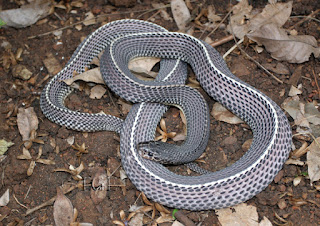

Comments
Post a Comment